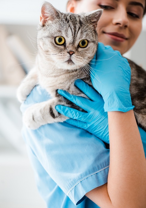
Abdominal Ultrasound​

Bladder, prostate, reproductive tract, intra-abdominal lymph nodes, kidneys, adrenals, spleen, stomach, pancreas, small & large intestine, liver, gallbladder & biliary tract, great vessels and mesenteric & serosal surfaces are evaluated.
​If warranted, ultrasound guided aspirates and/or biopsies can be performed subsequent to the initial scan. Please ensure that patients are fasted(NPO) for at least twelve hours prior to the scan with the exception of obligate drinkers. Gastrointestinal contents such as gas and ingesta can make the ultrasound exam very challenging and ultimately affect the diagnostic capabilities.
​Micro-convex, linear and convex probes are used alone or in combination in an effort to visualize all intra-abdominal structures. Even the largest patients can be scanned efficiently and with a high degree of sensitivity.
​Ultrasound examination for porto-systemic shunts is available.

​Thoracic Ultrasound

Every attempt will be made to evaluate the non-cardiac thoracic structures as well as the heart. Linear and micro-convex probes are used to provide as much detail as possible regarding the sternal lymph nodes, lung masses or consolidated lung tissue, parietal pleural masses, mediastinal masses and the pleural space. Please note that only lung lesions within the equatorial regions of the lung lobes are amenable to scanning.
​Thoracocentesis and/or ultrasound guided FNA or biopsy of thoracic masses can be performed subsequent to the initial scan. Please schedule these patients for the morning so that they can be observed over the day should they have an aspirate or biopsy procedure.

​Thyro-Parathyroid Complex and Cervical Ultrasound

Ultrasound of this complex is extremely helpful for those patients with unexplained hypercalcemia or a palpable cervical mass. In addition, by using a high frequency linear probe we are able to obtain detailed images of the salivary glands, the esophagus, great vessels, cervical musculature and the regional lymph nodes.

​Echocardiography

Foremost, echocardiography allows us to determine very quickly whether your patient’s clinical signs are attributable to cardiac disease. This will allow you to advance the treatment of your patient in a timely fashion and reduce unnecessary costs and therapies.

Once the problem has been determined to be cardiac in origin we can then evaluate or confirm the presence of:
  • Valve structure & motion (prolapse)
  • Chamber sizes
  • Systolic function (myocardial contractility)
  • Diastolic function
  • Wall thicknesses
  • Arrhythmia(s)
  • Pulmonary Hypertension
  • Congenital defects
  • Pericardial space
  • Heart base tumours
This information is collected through the use of two-dimensional and M-mode imaging and colour-flow, pulsed wave and continuous wave Doppler. We are happy to discuss the echocardiographic findings with you but also recognize the importance of veterinary cardiologists in the management of your patients. When appropriate we will recommend a second opinion by our cardiologist and assist in getting all the necessary information to them. Systemic blood pressure will be measured as part of the echocardiographic examination.

​Image and Report Management

After the examination is completed, all images and video clips will be saved in digital format and uploaded to a secure server so that they can be viewed and interpreted by our radiologist or cardiologist. Our scanning teams collaborate closely with the consultants to convey valuable history and technical impressions required to complete the presenting scenario. This process ensures a high level of accuracy and communication and maintains elite standards of quality care and control. Images in JPEG/AVI formats are available to all attending veterinarians wishing to discuss patient management and options with their clients.

​Interventional and Diagnostic Procedures

A compliment of ultrasound guided therepeutic and diagnostic procedures is available to all veterinarians. We will be sure to discuss the risks associated with this procedure and the alternatives available. Consultative process is available with our radiologist or cardiologist if required.
​We will have all of the necessary supplies to perform all of these diagnostic procedures. The cost for the materials is included in the aspirate charge. Once the sample is collected it will be the responsibility of that facility to prepare the sample for analysis and send in onward to a pathologist for study. A coagulation profile and direct blood smear evaluation for platelets is required prior to any biopsies and at the discretion of the attending veterinarian. Profiles are not required for fine needle aspirates. We would ask that clients be informed of this requirement and that they sign the appropriate consent form, available on the website in the “How to Book” section. Post procedure scanning will be performed to evaluate the site and adjacent organs. Twelve hour fasting (NPO) is also very important.
​ Sedation and/or anesthesia of the patient is the responsibility of the referring veterinarian. The staff of CMVUS will be happy to discuss options and also provide any local anesthesia that is required.